This is an archival copy of the Visualization Group's web page 1998 to 2017. For current information, please vist our group's new web page.
|
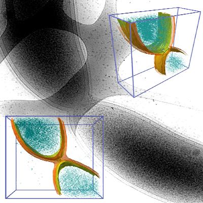
Bacteria contain a wealth of mechanisms that organize their internal and external structure. Electron Tomography offers the possibility to localize the cell components at a resolution that allows a better understanding of their structure and function. The 3D reconstruction of whole cells should lead to the ability to model spatial and temporal relationships among sub-cellular structures, organelles and macromolecular complexes.
Some of the three-dimensional reconstructions have a low signal to noise ratio resulting in data that produces difficult-to-interpret visualization output. Volume data with 2D features that are not easy to distinguish make a perfect case for construction of a model of structural features. The model - discrete representation of structure partially visible in noisy data - is easier to visually interpret, making it easier for researchers to understand the shape and topology of such features. In contrast, attempting to extract surfaces using isocontouring applied to noisy data produces results that are exteremely difficult if not impossible to interpret.
More information.
|
|