This is an archival copy of the Visualization Group's web page 1998 to 2017. For current information, please vist our group's new web page.
Toroidal Coiling of DNA - Comparing Observed Data with Computer Models

|
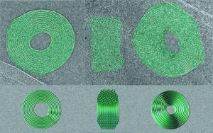
The figure shows three electron microscope images of DNA toroids accompanied by computer simulations of toroids in corresponding orientations. On the top left is a toroid seen nearly perpendicular to the direction of view, so that the ~23 Å spacing between rows of DNA strands, reflecting the hexagonal packing of strands throughout most of the toroid, is clearly seen around almost the entire toroid. The region where the fringe spacing is least visible may correspond to a region where the DNA strands cross over between loops at different radii. In the middle a toroid is seen edge on, revealing the hexagonal packing on the lower side and disordered packing on the top. The toroid in the right-hand image is tilted so that the fringes spacing is seen over only a small part of the toroid. Below each micrograph is a view of a solid model oriented to match the toroid. On the left, the region of crossovers is localized near the 4-o'clock position. The front half of the model in the center is cut away to reveal the hexagonal packing in the lower half and disorder in the crossover region toward the top.
The motivation for this work arises from the academic interest in the behavior of DNA, the fact that DNA is sometimes naturally packed in these toroidal arrays, for example in some sperm and bacteriophages, and the possibility that this may be a useful way to package DNA for genetic therapy.
This figure was created by Ken Downing, with assistance from the LBL Visualization Group on the high capacity visualization hardware located at the LBL Visualization Laboratory. The figure was submitted along with the manuscript entitled "Cryoelectron microscopy of lambda-phage DNA condensates" by N. V. Hud and K. H. Downing as a suggestion for the cover picture for the Proceedings of the National Academy of Sciences (PNAS).