This is an archival copy of the Visualization Group's web page 1998 to 2017. For current information, please vist our group's new web page.
Visualization of Theoretical and Observed Protein Molecules

|
Actin and tubulin are both protein molecules that polymerize into fibrous structures, known as the cytoskeleton, inside cells. Both types of fibers are involved in establishing cell shape, cell movement, and moving things around inside the cells.
The structure of actin was solved by a group of x-ray crystallographers in Ken Holmes' laboratory in Heidelberg (Kabsch, W., Mannherz, H. G., Suck, D., Pai, E. F., and Holmes, K. C. (1990) "Atomic structure of the actin:DNase I complex", Nature 347, 37-44 )
The tubulin structure was solved by Eva Nogales, Sharon Wolf and Kenneth Downing at Lawrence Berkeley National Lab (Nogales, E., Wolf, S. G., and Downing, K. H. (1998) "Structure of the alpha-beta tubulin dimer by electron crystallography", Nature 391, 199-203 )
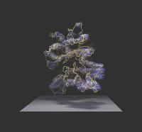
The "actin" and "tubulin-1 & -2" figures contain a tubular representation of the chain of amino acid subunits that make up the protein, inside two representations of the type of 3-D data that is used to determine the structure. In blue is a density map computed at medium resolution, while the density in yellow is calculated at a resolution high enough to indicate individual atoms. Both of these density maps were calculated from the atomic structure after it had been determined, in the process of checking validity of similar maps of other proteins determined at various resolution levels from experimental data.
The "tubulin-ribbon" figure shows a representation of the molecular structure that more clearly shows some of the types of sub-structures that the chain of subunits folds into - helices, sheets and loops - inside a density map computed that was generated from our experimental data at medium resolution. The structure of the protein was actually determined from a similar experimental map that had much higher resolution.
Also included in this figure is a ball-and-stick representation of a small molecule of GTP, an energy storage molecule, and a molecule of taxotere (similar to Bristol Meyers Squibb's trademark, Taxol). Taxol is an effective anti-cancer drug that stops cells from multiplying by binding to tubulin.
Among things one would like to do with supercomputers is to see what sort of internal flexibility exists in a molecule such as tubulin, and how binding of a drug like Taxol limits this flexibility. Information from such studies could point the way toward more effective drugs.
Ken Downing, UC Berkeley and LBNL.