Computer Vision Applications: Target Detection and Medical Images
Table of Contents
Introduction
Computer vision applications include steps as image processing, image analysis and classification, so that decision making based on images is feasible. We develop and apply many computer vision tools to medical micrographies and synthetic aperture radar images.1. Target detection: SAR image
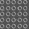 Fig.1.Front propagation to segment target from a 8-looks digital image. |
In collaboration with Prof. Regis Marques and Prof. Fatima Medeiros , we have designed a new framework for point target detection in synthetic aperture radar (SAR) images. We focus on the task of locating reflective small regions using a level set based algorithm. Unlike most of the approaches in image segmentation, we address an algorithm which incorporates speckle statistics instead of empirical parameters and also discards speckle filtering. The curve evolves according to speckle statistics, initially propagating with a maximum upward velocity in homogeneous areas. Our approach is validated by a series of tests on synthetic and real SAR images and compared with three other segmentation algorithms, demonstrating that it configures a novel and efficient method for target detection purpose. This research includes image analysis; object detection; partial differential equations; radar target recognition; speckle; synthetic aperture radar. |
Fig.2.Level set evolution driven by speckle statistics in synthetic aperture radar image: (a) propagation expansion speed function of a synthetic image with 8-looks speckle statistics, (b) arbitrary initial level set, (c) intermediary stage and (d) the final result.
2. Ocular Fundus
 Fig.6.Ocular fundus and vessel segmentation: (a) Segmentation result for Wavelet (b) Result of candidate extraction. |
Images of the ocular fundus presents indications about retinal, ophthalmic, and even systemic diseases such as diabetes, hypertension, and arteriosclerosis. Microaneurysms are the earliest sign of diabetic retinopathy and we aim at developing robust detection of microaneurysms in digital color fundus image as an auxiliar screening procedure. The current research involves the evaluation of a set of tools for vessel segmentation and how this task influences microaneurysms detection. We also propose a new approach to detect microaneurysms using mathematical morphology. Also, we have designed a pipeline for segmentation and the feature extraction to detect candidate microaneurysms. We show that the candidate microaneurysms detected with the proposed methodology have been successfully classified by a MLP neural network (correct classification of 84%). This work is a collaboration with LabVis group. |
Future Developments
1. Extend the research on target detection to oil spill and hurricane detection. 22. Run experiments in larger datasets to detect microaneurysms using mathematical morphology;
Publications
- R.C.P. Marques, F.N.S. Medeiros and D.M. Ushizima, "Target Detection in SAR Images Based on a Level Set Approach", IEEE Transactions on Systems, Man and Cybernetics - Applications, 2008. LBNL-958E.
- D.M. Ushizima and E. W. Bethel, "Pattern recognition on biomedical datasets", Imaging Science 2008, Annual Meeting of the Society for Industrial and Applied Mathematics. LBNL-1012E.
-
C.I.O. Martins, R.M.S.Veras, G.L.B.Ramalho, F.N.S.Medeiros, D.M. Ushizima, "Automatic Microaneurysm Detection and Characterization Through Digital Color Fundus Images",
Brazilian Artificial Intelligence Community Conference, Tenth Brazilian Symposium on Neural Networks, Proceedings of IEEE SBRN'2008, 2008. LBNL-.